IMAGINE
Imagine if a one-off treatment using personalised immunotherapy could fight cancer: CAR T-cell immunotherapy could make this a reality. Researchers at the University Hospital of Würzburg have been working on this for years. Now super-resolution microscopy is set to help optimise this special immunotherapy.
CAR T-cell immunotherapy is based on the body's own T-cells. These are cells of the immune system that recognise foreign structures and initiate a reaction to combat the foreign body. Unfortunately, T cells are not able to recognise tumour cells. However, they can be genetically engineered to recognise tumour cells and thus trigger a response from the immune system. This is exactly what happens in CAR T-cell immunotherapy: T-cells are taken from the patient and converted into CAR T-cells. They are then re-infused into the patient.
Every cell has certain structures on its surface, which are called antigens. T cells in turn have so-called antigen receptors on their cell surface, which they use to recognise these antigens. The lock-and-key principle ensures that receptors bind to the correct antigens: The structures fit together spatially like a key in a lock.

"Building" tumour cell-recognising T cells
CAR stands for chimeric antigenreceptors. Chimeras are known from Greek mythology as hybrid creatures and in biology refer to an organism that is made up of genetically different cells or tissues. A CAR is therefore an antigen receptor that is made up of different building blocks.
Firstly, however, the relevant antigens on the tumour cells to which the antigen receptors are to bind and, secondly, suitable T cells in the patient, from which CAR T cells are made, must be identified. T cells are then obtained from the patient's blood. In order to "incorporate" the CARs, a gene is introduced into the T cells using genetic engineering techniques. This gene ensures that the T cells can produce the antigen receptor themselves and incorporate it into the surface membrane. The CAR T cells are then returned to the patient by means of an infusion. Now they specifically recognise tumour cells in the body based on the antigen, can bind to them and destroy the tumour cell.
Ideally, a single treatment could therefore completely kill the tumour cells and even protect against recurrence for life, as the CAR-T cells remain in the body, multiply and form a memory.

For CAR T-cell therapy, T-cells are taken from the patient's blood, genetically converted into CAR T-cells and reintroduced into the patient via infusion. In the patient's body, the CAR-T cells specifically recognise tumour cells, can bind to them and kill them.
IMAGINE - a network of seven partners from science and industry
In future, the identification of tumour antigens and the characterisation of T cells are to be carried out using microscopic methods. The initiative project IMAGINE (Fighting Cancer with Optimal Personalised Immunotherapies) aims to help make this suitable for everyday use. The aim is to develop a super-resolution microscopy platform that can be used in everyday clinical practice. This will make it possible to identify the two target molecules with unprecedented precision, says project coordinator Prof Markus Sauer from the Chair of Biotechnology and Biophysics.
Super-resolution microscopy - insights into biological structures with almost molecular resolution
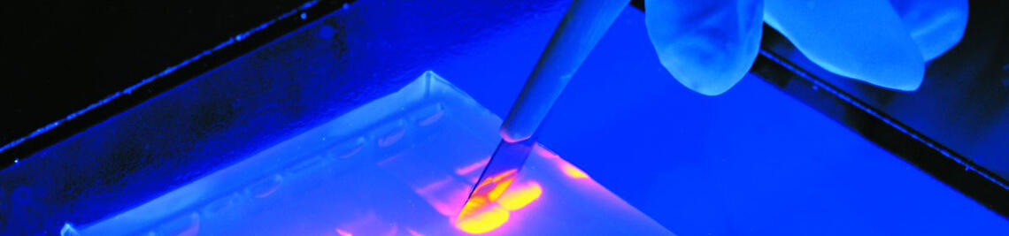
The limits of classical light microscopy are set by the physics of light. The diffraction limit will also be familiar to photographers who use apertures, aperture angles and lenses to take razor-sharp close-ups. Newer methodological approaches allow a much better resolution, without which many biological and medical insights would not have been gained. Super-resolution microscopy" is a collective term for various techniques such as STED, SIM and STORM, which use fluorescence microscopy to achieve resolutions beyond the diffraction limit. For comparison: in a conventional light microscope, two points less than 200 nm apart cannot be recognised as more than two separate points - with STORM, the resolution is approx. 20 nm.
Single-molecule localisation methods such as dSTORM(direct Stochastic Optical Reconstruction Microscopy) use fluorescent proteins that can switch between "on" and "off". The proteins therefore flash randomly and then switch off again. This prevents two neighbouring proteins from lighting up at the same time so that no more than two separate signals are detected. While the proteins flash alternately, the camera takes a series of images, from which computer calculations finally produce an image with extremely high resolution. It is assumed that each light spot represents a molecule, which is why the intensity of the fluorescence can be used to determine the number of molecules and their position.
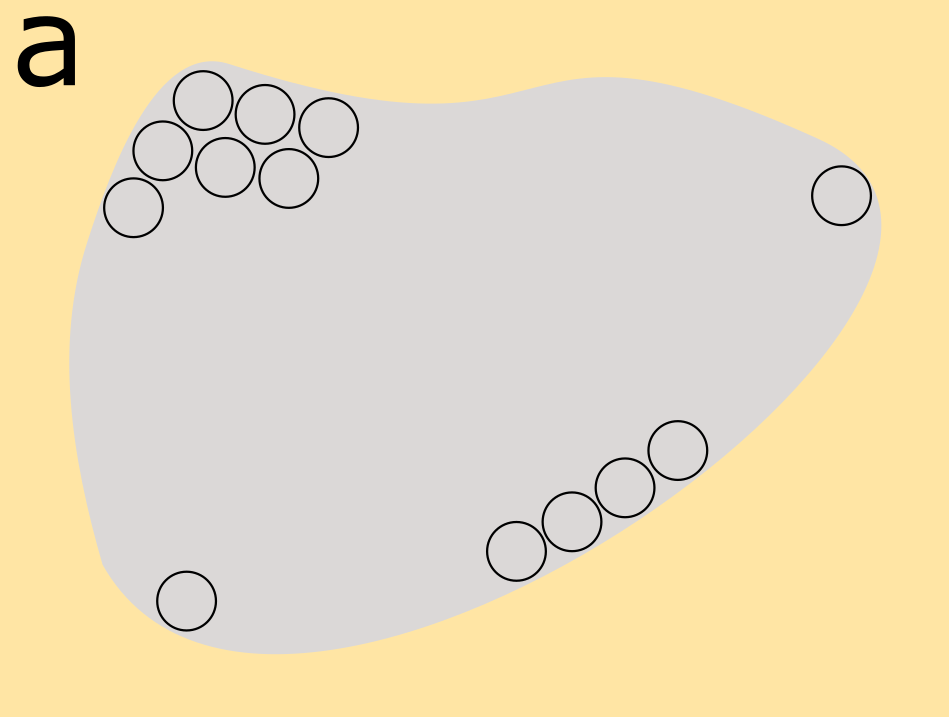
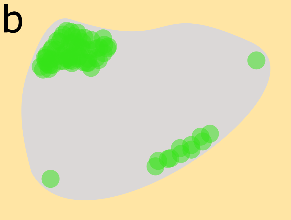
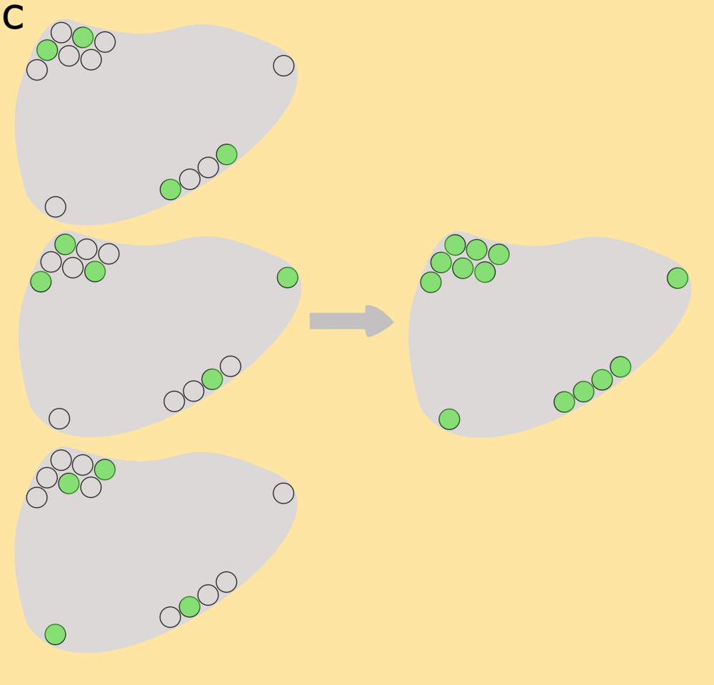
To visualise small structures in a cell, for example, you have to look at them one after the other. If proteins in close proximity fluoresce simultaneously, the resolution is no longer sufficient to detect individual points of light (b). The dSTORM techniqueuses flashing fluorophores: individual molecules flash alternately while the camera records thousands of images. Computer calculations ultimately lead to an image in which individual molecules can be visualised. (Images: Laura Colón/University of Würzburg)
The high resolution of the dSTORMmethod makes it possible to detect tumour cells that would remain undetected using conventional microscopy. 1000 antigens - i.e. 1000 individual molecules - would be required to detect a tumour cell using standard methods. In contrast, a very small amount of antigens is sufficient to detect corresponding antigens with ddSTORM. "This means that cells can also be recognised that were previously undetectable by diagnostic means and therefore significantly more patients can be identified who can be successfully treated with immunotherapy," says Prof. Sauer.

CAR-T cell research for 10 years at the University Hospital of Würzburg
Also involved in the project is a team led by Clinic Director Prof Dr Hermann Einsele, Prof Dr Michael Hudecek and Dr Thomas Nerreter from the Medical Clinic and Polyclinic II of the University Hospital of Würzburg (UKW). The UKW has been researching CAR-T cells for 10 years: Michael Hudecek, who had previously conducted research on CAR-T cells in the USA, came to Würzburg in 2012. Based on his experience, a research laboratory and a working group were established at the UKW, which today conduct cutting-edge research at an international level.
The team also has experience in the preclinical development and clinical application of the first preparations, including for leukaemia. Patients very often respond to CAR T-cell therapy, but often suffer a relapse, explains Prof Dr Einsele. The joint project aims to improve existing treatment options and accelerate the development of treatments for other cancers.
Immunotherapies have great potential for use in other diseases such as infectious diseases, autoimmune diseases, degenerative diseases and cardiovascular diseases.
The project is funded by the Federal Ministry of Education and Research (BMBF). The project started on 1 October 2021 and will run for three years.
Project partners
- Chair of Biotechnology and Biophysics, Julius-Maximilians-Universität Würzburg (coordination)
- Medical Clinic and Polyclinic II, University Hospital Würzburg
- Miltenyi Biotec B.V. & Co. KG, Bergisch Gladbach
- LaVision BioTec GmbH, Bielefeld
- Max Planck Society for the Advancement of Science e.V. - Max Planck Institute of Biochemistry, Planegg
- Massive Photonics GmbH, Gräfelfing
- T-CURX GmbH, Würzburg
Contact us
Project coordinator Prof Dr Markus Sauer, Chair of Biotechnology and Biophysics, Julius-Maximilians-Universität Würzburg,m.sauer@uni-wuerzburg.de
Press release:
https://www.uni-wuerzburg.de/aktuelles/pressemitteilungen/single/news/neuer-schub-fuer-immuntherapien-1/
Hudecek Lab:
https://www.ukw.de/medizinische-klinik-ii/forschung/t-zell-engineering/
https://www.ukw.de/research/research-hudecek-lab/home/
Chair of Biotechnology and Biophysics
https://www.biozentrum.uni-wuerzburg.de/super-resolution/