Plant Signaling Pathways Decoded
08/28/2024Using newly generated “optogenetic” tobacco plants, research teams from the University of Würzburg's Departments of Plant Physiology and Neurophysiology have investigated how plants process external signals.

When it comes to survival, plants have a huge disadvantage compared to many other living organisms: they cannot simply change their location if predators or pathogens attack them or the environmental conditions change to their disadvantage.
For this reason, plants have developed different strategies with which they react to such attacks. Such reactions are usually triggered by certain signals from the environment. As has long been known, the intracellular calcium concentration plays an important role in the processing of these signals.
However, in addition to changes in the cytoplasmic calcium level, changes in the cell's membrane potential have also been suspected as a signal transmitter. Research groups from the Departments of Neurophysiology, Pharmaceutical Biology and Botany at Julius-Maximilians-Universität Würzburg (JMU) have investigated the calcium-membrane potential relationship in more detail. They have now published their findings in the journal Nature.
Light-sensitive Channels Enable Targeted Manipulations
For their study, the research teams worked with tobacco plants that carry ion channels that can be specifically switched on with light. More than 20 years ago, Peter Hegemann, Georg Nagel and Ernst Bamberg initiated the success of optogenetics, with their discovery and characterization of light-activated ion channels, so-called channelrhodopsins. With the help of these light-sensitive proteins, which are obtained from algae and microorganisms, the JMU researchers were able to experimentally investigate whether the influx of calcium ions or anion efflux-mediated depolarization of the cell membrane is decisive for the plant's reaction to a certain stress situation. However, the scientists had to do a great deal of preparatory work before they were able to do this.
Optogenetics with Rhodopsins
Channelrhodopsins, ion channels that carry an intrinsic rhodopsin-based light switch, revolutionized neuroscience through the light-controlled investigation of neuronal networks. The use of channelrhodopsins in plant research only became possible 20 years later, through close collaboration between the group of Georg Nagel, Professor at the Institute of Physiology at JMU, and plant researchers from the Würzburg Chairs of Botany 1, 2 and Pharmaceutical Biology.
In 2021, Georg Nagel's group, together with Dr. Kai Konrad, group leader at the JMU Chair of Prof. Rainer Hedrich Botany 1, published an approach to optimize the use of channelrhodopsins in plants by overcoming three main difficulties.
Rhodopsins Require Vitamin A
Point 1: “Like all rhodopsins, including those in our eyes, channelrhodopsins require the small molecule retinal, also known as vitamin A, to absorb light. We humans get retinal mainly from beta-carotene, the provitamin A. However, land plants do not contain retinal, but a lot of beta-carotene,” explains Dr. Shiqiang Gao, co-author of the Nature publication and ‘rhodopsin engineer’ from the Optogenetics lab of the Department of Neurophysiology at JMU.
In 2021, Gao succeeded for the first time in combining the expression of channelrhodopsins with the production of retinal from beta-carotene in plant cells. This enabled the development of tobacco plants with a high retinal content and successful expression of channelrhodopsins.
Dr. Markus Krischke from the Metabolomics Core Unit at the Department of Pharmaceutical Biology headed by Professor Martin Müller at JMU Würzburg confirmed the high retinal content of the various transgenic tobacco plants.
Comparable transgenic tobacco plants were produced for the recently published study by Dr. Meiqi Ding from the Department of Botany I under the direction of plant physiologist and expert for plant signal processing Dr. Kai Konrad from the group of Professor Rainer Hedrich at the Department of Botany I.
Plants Need Light to Grow
Point 2: “Most rhodopsins are activated by blue or green light. However, this is always a component of white light,” explains Georg Nagel. As a result, the tobacco plants could not be grown in a greenhouse or under artificial white light, as is usually the case. Only in special growth chambers with red LED light, which can be used photosynthetically, it was possible to avoid unwanted rhodopsin activation. Tests under different growth conditions showed: “Tobacco develops healthily and unchanged under red light compared to greenhouse conditions,” says Dr. Kai Konrad.
Functional Expression of Channelrhodopsins in Plants
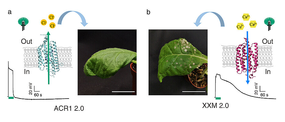
Point 3: The expression of chanelrhodopsin in tobacco cells often causes difficulties. In 2021, the Würzburg team of scientists succeeded in expressing the light-activated anion channel GtACR1 in tobacco plant cells. As a result, Georg Nagel's team was able to develop various channelrhodopsins that were optimized for the permeability of calcium ions. Finally, Dr. Shiqiang Gao and Dr. Shang Yang, both members of Nagel's group, succeeded in developing a very good calcium-conducting channelrhodopsin XXM 2.0 for targeted expression in tobacco plants.
This was the breakthrough: “The successful expression of channelrhodopsins with different ion selectivity in plant cells enables the comparison of different ion signals in parallel to the electrical signal, the so-called depolarization,” explains Dr. Meiqi Ding. She used the calcium-conducting channelrhodopsin XXM 2.0 and the light-activated anion channel GtACR1 to investigate the different ion signaling pathways in tobacco.
A New Era in Plant Research
These newly generated “optogenetic” tobacco plants made it possible to clarify the question of whether calcium influx or membrane depolarization is decisive for the plant's response to a specific stress situation. “The answer was clear,” says Dr. Kai Konrad, corresponding author. First author Dr. Meiqi Ding from Dr. Konrad's group explains, “After activation of the anion channel, the leaves wilted and responded with the typical plant response to drought; the plant hormone abscisic acid (ABA) was produced and gene expression was ramped up to protect against desiccation.”
“However, in the plants with the calcium channel, there was no change in ABA levels after optogenetic stimulation,” Dr. Ding continued. “Instead, the plants produced signal molecules and plant hormones to initiate defence mechanisms against predators, recognizable by white spots on the leaves,” said Dr. Konrad.
Dr Sönke Scherzer at the chair of Prof Hedrich was able to show by direct ROS measurements that reactive oxygen species (ROS) are released in the process.
Dirk Becker and Rainer Hedrich at the Chair of Botany 1, designed an experimental approach to support the working hypothesis using transcriptomic and bioinformatic analysis.
The scientists are convinced that this study is just the beginning of a new era in plant research. Ultimately, the signaling pathways of plants can now be better “illuminated” using various rhodopsins.
Original Publication
Probing plant signal processing optogenetically by two channelrhodopsins. Meiqi Ding, Yang Zhou, Dirk Becker, Shang Yang, Markus Krischke, Sönke Scherzer, Jing Yu-Strzelczyk, Martin J. Mueller, Rainer Hedrich, Georg Nagel, Shiqiang Gao, Kai R. Konrad. Nature, 28 August 2024. DOI: 10.1038/s41586-024-07884-1. https://www.nature.com/articles/s41586-024-07884-1
Contact
Dr. Kai Konrad, Julius-Maximilians-Universität Würzburg, kai.konrad@uni-wuerzburg.de
Dr. Shiqiang Gao, Institute of Physiology – Neurophysiology, gao.shiqiang@uni-wuerzburg.de


