Continuously Pushing the Boundaries of Eye Research
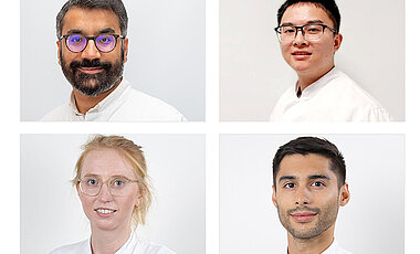
10/07/2025A research team from the Würzburg Eye Clinic led by Dr. Malik Salman Haider celebrates success at the congress of the German Ophthalmological Society.
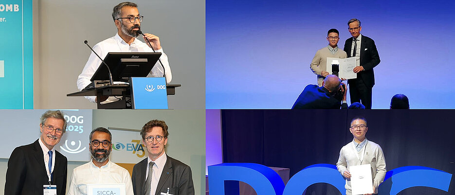
The congress of the German Ophthalmological Society (DOG), which took place in Berlin from 25 to 28 September, had the guiding theme "Ophthalmology in transition - shaping the future together" this year. The research team from the Department of Ophthalmology at the University Hospital of Würzburg once again presented numerous innovations that are changing ophthalmology in the long term and inspiring new therapeutic approaches.
"As head of our research team, I am incredibly proud of the outstanding achievements that we presented at DOG 2025. From the first 3D conjunctival spheroids to spheroid-based microtissues of the meibomian glands and innovative degradable hydrogels with dexamethasone - our team is continuously pushing the boundaries of eye research," says Dr. Malik Haider. "Our work is more than just science - it is a commitment to developing reproducible, patient-centred and translational solutions."
Best Abstract Award to Zhi Liang for the Biofabrication of Three-dimensional Conjunctival Spheroids as an In-vitro Test System
Numerous papers were honoured at the congress. Zhi Liang received the "Best Abstract" prize of the AG Young DOG. The prize, endowed with 500 euros and donated by Margarete Kramer, recognises outstanding scientific work by young ophthalmologists and scientists from the entire field of ophthalmology. At the congress, Zhi Liang presented the first three-dimensional conjunctival spheroid model, which addresses the lack of physiologically relevant in-vitro systems for ocular surface research.
Using special honeycomb-shaped cell culture plates made from agarose - a sugar building block derived from red algae - and human conjunctival cells, Zhi Liang and his team were able to produce small spherical mini-tissues known as spheroids. These consist of several layers of cells, similar to the real conjunctiva of the eye. "Our reproducible and cost-effective platform promotes mechanistic studies and the screening of active substances, complies with the 3R principles and offers potential for personalised eye treatments," explains Malik Haider.
Sicca Prize for Biofabrication of Spheroid-based Microtissues from Meibomian and Lacrimal Glands
Malik Haider himself was honoured with the Sicca Prize, which is endowed with 1,500 euros. He also presented a paper from the field of biofabrication. In order to counteract the lack of physiologically relevant models for research into dry eye diseases and the administration of eye medication, he developed spheroid-based microtissue from meibomian and lacrimal glands. Meibomian glands are small sebaceous glands at the edge of the eyelids. They produce an oily fluid that forms a thin layer on the tear film and thus prevents the tear fluid from evaporating too quickly. As lipid suppliers of the tear film, they are crucial for healthy, moisturised eyes.
Using scalable 3D culture methods, Haider and his team succeeded in getting the cells to coalesce into uniform, viable, spherical mini-tissues. These retained the tissue-specific structure and function of the real glandular tissue. The method is simple, inexpensive and easily reproducible. This makes it ideal for researching the causes of diseases, testing new therapies and even for personalised ophthalmology. It also fulfils the 3R principles as it can significantly reduce animal testing.
Two Posters of the Day
In addition, two poster contributions from the Würzburg team were honoured as Poster of the Day. Dr. Raoul Verma-Führing was honoured for the development and characterisation of a novel carrier system for ophthalmic drugs. This is a hyaluronic acid-based gel filled with tiny micelles. These contain the active ingredient dexamethasone, a powerful anti-inflammatory drug. The system showed excellent cytocompatibility and is therefore cell-compatible and safe for the eye.
It has stable swelling behaviour, meaning it can absorb water without losing its stability. It can also be adjusted so that it degrades slowly in the eye and releases the active ingredient evenly and over a longer period of time. The combination of light activation during insertion and the sensitive reaction to the body's own enzymes creates a customised and patient-friendly method for the effective treatment of eyes over longer periods of time - without the need for constant new drops or injections.
The second poster of the day from Würzburg was presented by Pia Schröder. In her work, the assistant doctor drew attention to the discrepancy between ganglion cell analysis in optical coherence tomography (OCT) and visual field tests in the detection of (post-)chiasmatic lesions. The chiasm is the junction where the optic nerves that transmit signals from the eye to the brain meet. Damage or disease at or behind this junction can lead to visual field loss. Glaucoma can also damage the optic nerves and cause visual field loss.
It can be difficult to distinguish damage to the optic nerve caused by glaucoma from damage behind the optic nerve junction in the brain. The nerve cell layer of the retina can be analysed in great detail using a special imaging method, OCT ganglion cell analysis. Pia Schröder reported on three patients in whom this analysis showed a thinning of the nerve cell layer along the vertical centre line, although the usual visual field tests were still unremarkable. Further examinations revealed a tumour of the pituitary gland (pituitary adenoma), scarring along the optic nerve pathway (gliosis) and a lesion in the area of the capsular saddle/hypothalamus. The results show that changes in the ganglion cell layer can occur at a very early stage, even before patients notice restrictions in the visual field. OCT analysis could therefore be an important early warning sign.
Malik Haider summarises: "These awards and recognition reflect not only the individual brilliance, but also the collective commitment, creativity and perseverance of each individual team member."


