

Dentistry: Improved Diagnosis of Crown-root Fractures
11/18/2025A study from the Würzburg University Hospital provides practical findings for the management of tooth fractures following an accident. Dr Ralf Krug has now received an award for his work.
Treatment is usually very complex for teeth that are broken from the visible tooth crown deep into the tooth root. Smaller, loosened pieces of broken tooth are often also present. Such crown-root fractures usually affect children, adolescents and young adults who have had an accident.
After reattachment of tooth fragments, there are often complications that reduce the long-term results of the treatment. This was shown in a 2020 study by the Polyclinic for Tooth Preservation and Periodontology and the Dental Accident Centre at the University Hospital of Würzburg (UKW).
In another study from 2024, the Würzburg research group examined various tooth-preserving techniques and found, among other things, a complication rate of 19 per cent after 3.6 years.
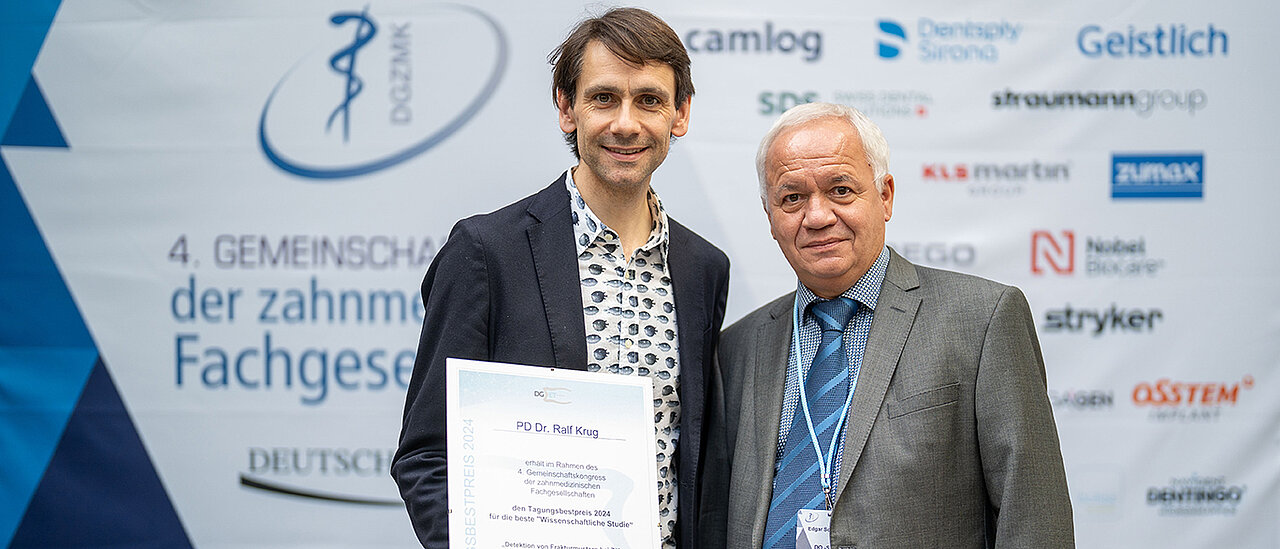
Award Ceremony at Congress in Berlin
How well can the exact location and depth of crown-root fractures be determined using various examination methods? Which of the resulting findings are important for treatment? To find out, Dr Ralf Krug, senior consultant and specialist in endodontology at the UKW, analysed the findings of primary diagnostics in more detail.
He presented his findings at the annual conference of the German Society for Endodontology and Dental Traumatology in Hamburg in 2024. He was awarded the Tagungsbestpreis for his presentation at this year's congress, which took place in Berlin at the end of October 2025.
For the study, Dr Krug examined 28 patients with 38 crown-root fractures, predominantly on incisors. He carried out a thorough clinical examination with photos, X-rays and a special high-resolution digital volume tomography (DVT) in 3D. This made it possible to assess features such as the exact location of the fracture, the depth of the fracture, fine cracks and additional fractures in the root or jawbone.
3D Images Improve Primary Diagnostics
"Our results show that 3D images can be particularly helpful during the initial examination in order to assess the necessary depth of the fracture from a tooth-preserving perspective and to rule out any additional root fractures," says Ralf Krug. Before treatment, a thorough clinical inspection of the fracture surface is essential in order to recognise the finest cracks and partial fragments.
As dental implants cannot be placed in children, adolescents and young adults due to their age, it makes sense to assess the damaged teeth as reliably as possible before treatment: "Our data shows that with precise primary diagnostics, teeth with crown-root fractures can be preserved in a stable and long-term manner in many cases," says Ralf Krug.
His project was funded as part of a DGR²Z-GC grant from the German Society for Restorative and Regenerative Dentistry in cooperation with the dental company GC Germany GmbH.
Publications
Soliman et al. 2020 found up to 56.8 % (25/44) restorative and 22.7 % (10/44) endodontic complications for adhesive fragment luting therapy. The functional survival of the refixed fragments was 66.7 % (26/39) after ⌀ 9.5 years. Soliman S, Lang LM, Hahn B, Reich S, Schlagenhauf U, Krastl G, Krug R. Long-term outcome of adhesive fragment reattachment in crown-root fractured teeth. Dent Traumatol. 2020 Aug;36(4):417-426. https://doi.org/10.1111/edt.12550
Krug R, Soliman S, Reich S, Winkler A, Hahn B, Krastl G. Periodontal status after treatment of teeth with crown-root fracture - a prospective observational study. Joint annual conference DGZ & DGPRO, 13-15 June 2024, Leipzig (short presentation, PDF)
Krug R, Soliman S, Connert T, Kroeger AT, Dietrich T, Krastl G. Detection of fracture patterns in teeth with crown-root fracture in primary care. 13th Annual Conference DGET, 21-23 November 2024, Hamburg (short presentation, PDF)


