

Pain subsides depending on gender
07/07/2026How pain subsides depends on gender – even though the immediate reaction to the injury is largely the same. Researchers at Würzburg University Hospital have made this observation in an animal model.
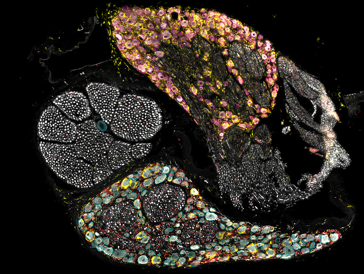
“We already have a fairly good understanding of what pain is and how it arises. But how does it subside again? What exactly happens in the central hub – the spinal ganglion – where pain signals from the injured peripheral nerves are received and relayed via the spinal cord to the brain?” asks Professor Robert Blum.
The molecular and cell biologist at the Department of Neurology and Polyclinic of the University Hospital of Würzburg (UKW), has, together with his research group as part of the KFO5001 Clinical Research Group, taken a closer look at this key site for pain processing, also known as the DRG (dorsal root ganglion). Specifically, his team carried out a comprehensive analysis of the molecular and cellular levels in the healthy state, during the acute pain phase and during the so-called resolution phase – that is, the phase in which the pain has naturally been reduced by at least half.
AI-assisted analysis
For their study, the researchers used an established rat model for neuropathic pain. The research group has been investigating this model for many years in collaboration with Professor Heike Rittner, holder of the Chair of Pain Medicine, and Professor Alexander Brack from the Department of Anaesthesiology. In this model, the sciatic nerve is injured. Unlike in many other models, however, the signs of pain subside over time.
Seven days as well as five weeks after the nerve injury, the researchers harvested the DRGs. Each ganglion was dissected into more than 100 wafer-thin tissue sections, mounted on microscope slides and stained with antibodies that specifically visualise nerve cells, satellite glial cells and macrophages – the ‘scavenger cells’ of the immune system. The specimens were then imaged under a microscope.
“In total, we had around 7,500 four-channel microscopy images, which amounts to roughly 30,000 individual images of the different cell types. No human being can evaluate these objectively,” said Blum. This is where an AI application came into play, which the research group had specifically trained for the analysis of the DRG. The method is based on deep learning. In this process, artificial neural networks are trained on image characteristics so that they can subsequently evaluate a large number of images objectively.
In this way, the group was able to produce, for the first time, an overall picture of how the cells surrounding the neurons are distributed during the phases when pain is at its most intense or is subsiding. Surprisingly, contrary to long-held assumptions, the nerve cells in the DRG are not eliminated but remain largely intact. Instead, it is primarily the cell activity and the spatial arrangement of the surrounding cells that undergo dynamic changes – and these differ between female and male animals.
Macrophages migrate towards the neurons during the pain phase
For example, during the acute pain phase, macrophages release inflammatory mediators which make the so-called nociceptors more responsive to stimuli. This increases pain perception. Initially, the macrophages migrate close to the surface of the neurons, displacing the satellite glial cells that normally reside there. When the pain subsides, the immune cells migrate out of this intercellular space again.
Many assume that the satellite glial cells also reprogramme themselves into a kind of macrophage-like immune cell. “However, we can demonstrate in high resolution that these are two different cell types. During the pain phase, the macrophages migrate between the neuron and the satellite glial cells and then back out again. Anyone can see this for themselves,” says Dr Annemarie Sodmann. The neurobiologist and co-author is an expert in the analysis of complex biological data. She has visualised the images in a publicly available Streamlit app.
In females, the macrophages remain active for longer
Another surprise emerged from the gender-specific analysis. It appears that males and females process pain differently at a biological level. In male rats, the macrophages are slightly more flexible and disappear more quickly from the surface of the neurons. In female rats, by contrast, the macrophages were still clearly visible on the neurons even after five weeks.
This suggests that the immune response appears to remain active for longer in females. It is interesting to note, however, that both sexes reach the same endpoint: the pain subsides at the same point in time, albeit following different patterns.
In males, contact between satellite glial cells and neurons persists for longer
For lead author Felicitas Schlott, the clear difference in the glial response was the biggest surprise. In the DRG, within the peripheral nervous system, satellite glial cells envelop the cell bodies of sensory neurons like a ‘sheath’ and influence their activity. This means they can amplify pain signals. During the acute pain phase, the activation marker for satellite glial cells (GFAP) was similarly high in both male and female animals. In males, however, the activation persisted for longer.
“Although there were already indications that immune parameters are regulated differently in the two sexes, this pronounced inverse reaction between macrophages and satellite glial cells is extremely fascinating,” says Schlott. The project was also the subject of her PhD thesis, which she recently successfully defended.
Specific genes confirm sex-specific programmes
To provide further evidence, the team, in collaboration with the Core Unit Systems Medicine at the University of Würzburg’s Faculty of Medicine and the Interdisciplinary UKW Centre for Clinical Research (IZKF), sequenced the complete transcriptome – that is, the genes expressed throughout the DRG tissue. “The central mechanisms of pain processing are evolutionarily conserved and are therefore very similar in both sexes. We assume that the observed differences in pain resolution are attributable to subtle biological differences, such as the sex-specific distribution of cells within the DRG or the regulation of certain gene groups,” explains Blum.
During the acute pain phase, the genes that were massively up-regulated during pain development were largely similar between the sexes. However, during pain resolution, these overlaps decreased and new, relevant genes emerged.
“I wouldn’t have thought that individual genes would go through the roof like that,” says Blum. “This suggests that pain resolution is not a reversal of the injury – in other words, not a return to the original state – but an entirely new genetic and sex-specific programme.”
Overall, according to Schlott, the study shows that sex plays a much greater role in cellular and molecular pain biology than previously assumed. “However, the very fundamental biological causes of these differences have been far too little investigated, both in humans and in animals.”
Implications for future personalised pain medicine
For pain research, the findings published in the journal Cell Reports could represent a step towards taking a broader view of sex-specific differences in pain processing. This is because the review shows that pain resolution is a multicellular, multicategorical and sex-specific process that needs to be interpreted.
Blum describes the biological processes involved in pain resolution as a self-optimising, perhaps chaotic oscillation of many factors, all of which lead to a single goal. He compares this process to a swarm of bees, in which the flight pattern of an individual bee may appear chaotic, yet all the bees fly in the same direction. It therefore makes no sense to single out one bee or one factor and eliminate it in order to accelerate the resolution of pain.
The next step is to investigate whether the changes in the DRG described in the study can also be applied to humans. To this end, systemic parameters from blood samples taken from humans will be compared with those from the animal models. A further step that Annemarie Sodmann plans to take by setting up her own research group is to apply these new findings to regenerative medicine: can the resolution of neuropathic pain be promoted by repairing nerve damage? And does this also trigger the gender-specific genetic and cellular programmes involved in pain regression?
Publication
Felicitas Schlott, Beate Hartmannsberger, Thorsten Bischler, Tom Gräfenhan, Alexander Brack, Heike L. Rittner, Annemarie Sodmann, Robert Blum. Sex-specific molecular and cellular phenotypes during resolution of neuropathic pain in dorsal root ganglia. Cell Reports. Volume 45, Issue 6, 2026, 117442, ISSN 2211-1247, https://doi.org/10.1016/j.celrep.2026.117442