

Exploring new treatments for the eye using artificial tissue
10/06/2026The Department of Ophthalmology at Würzburg University Hospital was honoured at the congress of the German Ophthalmological Society for a new conjunctival model.
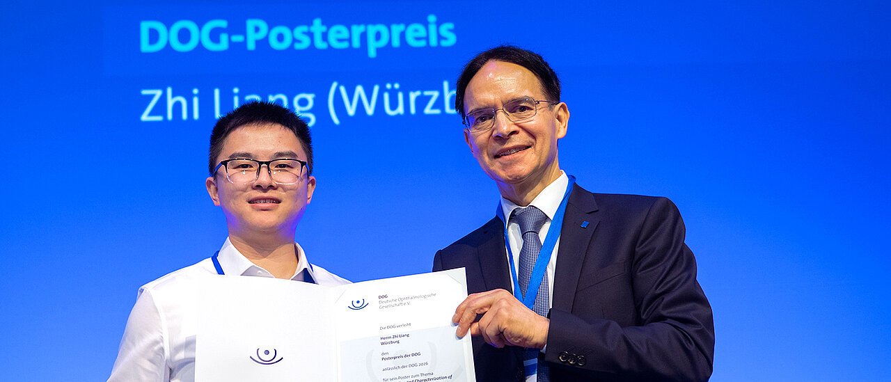
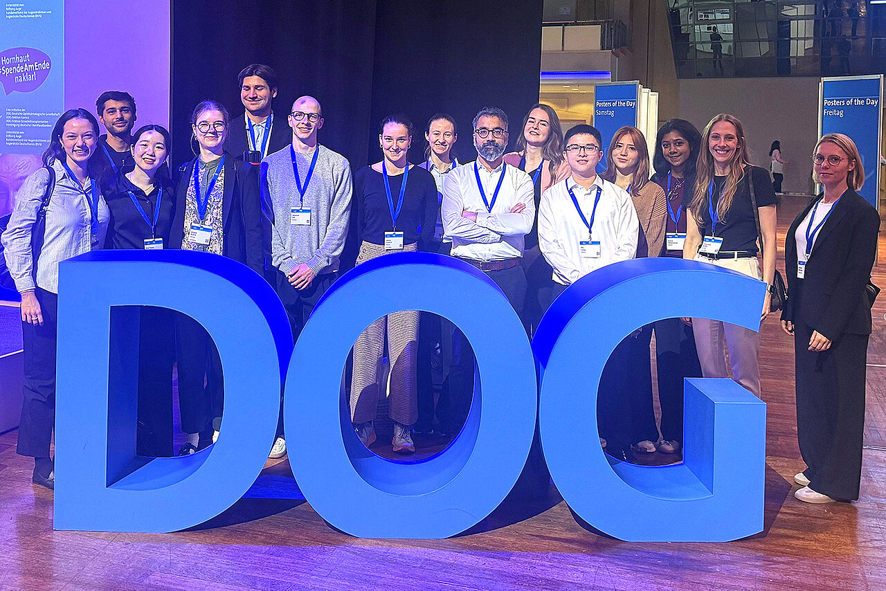
How can human tissue be replicated as realistically as possible? And how can such models help to develop new treatments for eye diseases and reduce the need for animal testing? These are the questions being addressed by the research team led by Dr Malik Salman Haider at the Department of Ophthalmology at Würzburg University Hospital (UKW), under the direction of Professor Jost Hillenkamp. At this year’s congress of the German Ophthalmological Society (DOG) in Berlin, the team’s work was awarded a poster prize.
Producing conjunctival tissue in the laboratory
At the heart of the award-winning work, presented by PhD student Zhi Liang, is a newly developed scaffold for producing conjunctival tissue. The conjunctiva protects the sensitive surface of the eye and helps to keep it moist and resilient. However, tissue can be lost following injuries, inflammation or surgery. Realistic models of the conjunctiva are therefore needed for the development of tissue replacements and new treatment options.
The project is the result of close and fruitful collaboration with the Chair of Functional Materials in Medicine and Dentistry (FMZ), headed by Professor Jürgen Groll. Wahaj ul Haq from the Eye Clinic, Dr Camilla Mussoni and Muhammad Aslam from the FMZ, and Zhi Liang from the Eye Clinic jointly developed a precisely structured scaffold. It features clearly distinct upper and lower regions, which enable epithelial cells and fibroblasts to be organised in accordance with their natural arrangement. This results in a tissue-like, two-layered construct.
“I am very proud of the whole team. This award reflects the hard work, creativity and perseverance of our young researchers, whose commitment drives our research forward,” said Malik Salman Haider, commenting on the success. “I am particularly pleased that their work is being recognised at such a prestigious conference.”
The platform provides a basis for the further development of a tissue substitute for the conjunctiva and of standardised three-dimensional models of the ocular surface. It could also support the investigation of new treatment options and drug delivery systems, whilst helping to replace and reduce animal testing.
Start-up funding for research into fungal keratitis
In addition to the poster prize, the research team received DOG seed funding of 2,500 euros in connection with the clinic’s DFG-funded project on fungal keratitis. In fungal keratitis, fungi infect the cornea and can cause severe tissue damage. In the worst-case scenario, this can lead to loss of vision. The aim of the funded project is to develop new ways of delivering medication directly to the affected area. More effective drug delivery could help to improve the treatment of fungal infections of the cornea.
This year’s DOG Congress, which took place in Berlin from 24 to 27 September, brought together more than 3,100 specialist delegates and a total of more than 4,600 participants.
Additional images