
New approaches for treatment of brain metastases after breast cancer
02/24/2026A novel 3D model recreates the environment of nerve cells in the brain and shows how the neuronal network changes due to brain metastases after breast cancer. It was developed by researchers at the University Hospital of Würzburg.
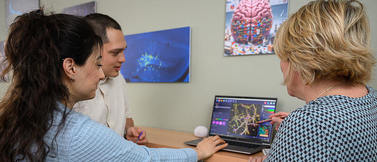
Brain metastases in breast cancer are difficult to treat and are associated with a poor prognosis. A current study by Esra Türker and Mateo Andrade Mier provides new insights into the question of the cellular partners of the tumour cells and their interaction with the surrounding brain tissue.
Both researchers are part of Professor Carmen Villmann's research group at the Institute of Clinical Neurobiology at the University Hospital of Würzburg (UKW). The results have been published in the journal Advanced Functional Materials.
Carmen Villmann's team developed a 3D cell culture model based on a special hydrogel that contains important components of the extracellular matrix in the brain, as well as structure-giving scaffolds. The various cell types are introduced into these scaffolds. HER2-positive breast cancer cells, which have a particularly high risk of brain metastases, were investigated.
Tumour cells from the breast adapt to the new environment in the brain
The results show that the tumour cells originating from the breast adapt to their new environment in the brain and form close contacts with nerve cells and astrocytes. Among other things, the latter form the blood-brain barrier.
The activity of the nerve cells increases significantly. Electron microscopic analyses indicate that synapse-like contact points develop between tumour cells and brain cells, via which growth-promoting signals could be transmitted.
These findings open up new therapeutic strategies: in future, treatments could be aimed at first interrupting these cell contacts and only then specifically destroying the tumour cells. The established 3D model is also suitable for systematic drug testing and can be transferred to other types of tumours with brain metastases.
Publication
Esra Türker, Mateo S. Andrade Mier, Jessica Faber, Mike Friedrich, Zan Lamberger, Jeannette Weigelt, Christian Stigloher, Nicoletta Murenu, Natascha Schaefer, Jörg Tessmar, Gregor Lang, Silvia Budday, Katrin G. Heinze, Antje Appelt-Menzel, Pamela L. Strissel, Reiner Strick, Carmen Villmann. A 3D Biofabricated Disease Model Mimicking the Brain Extracellular Matrix Suitable to Characterize Intrinsic Neuronal Network Alterations in the Presence of a Breast Tumor Disseminated to the Brain. Advanced Functional Materials. First published: 27 November 2025. https://doi.org/10.1002/adfm.202515220