
Medicine: A student's journey into brain research
07/07/2026A brain atlas designed to make micrometre-scale anatomy usable in everyday clinical practice. Helen Friedrich, a medical student at the University of Würzburg, is working on that. A conversation about her path into research.
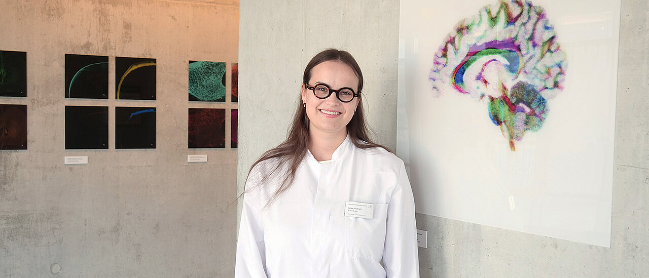
Helen Friedrich is studying medicine in Würzburg and conducting research at the interface between neuroanatomy, imaging and clinical medicine. Her PhD research focuses on high-resolution brain imaging and the question of how anatomical knowledge can be put to use for neurological patients.
Using an exceptionally high-resolution 7-Tesla MRI scan as a basis, she has worked with an interdisciplinary team to create a detailed MRI atlas of brain structures. As routine clinical cranial MRI scans have a significantly lower spatial resolution, she has converted this atlas into a standardised reference space.
This means that, in future, high-resolution anatomical information can also be used for clinical analyses. For her research, Helen Friedrich spent more than one and a half years at Brigham and Women’s Hospital, Harvard Medical School in Boston, USA.
Helen, your career path has taken you across various disciplines. What drives you?
Helen Friedrich: People. I initially studied law, whilst taking a particular interest in questions of legal philosophy and ethics: What is justice? How do we make decisions? How do we reflect on responsibility and human life? These questions led me to psychology. Whilst studying for my bachelor’s degree in Würzburg, I developed a keen fascination for functional magnetic resonance imaging.
This method uses magnetic fields to detect signals from the brain and visualise brain functions. The possibility of investigating human thought and perception processes using imaging techniques made a lasting impression on me. As part of my bachelor’s thesis under Professor Matthias Gamer, I collaborated on a study into social attention using functional magnetic resonance imaging.
How did you come to switch to medicine?
I think it was curiosity and perseverance. Whilst working part-time in neuropsychology, I encountered a patient who developed a prolonged state of confusion following an operation. This was accompanied by dizziness, a tremor, anxiety and a complex psychopharmacological profile. I realised at the time that, on the one hand, I lacked the medical knowledge to properly classify these symptoms and, on the other, the clinical expertise to investigate such a case.
I did not want to leave this gap unaddressed. I wanted to understand what lies behind such clinical courses: what can be explained by the operation, what role medication plays, and what changes in brain function account for these neurological phenomena. This need gave rise to my desire to study medicine. Whilst studying for my Master’s in Psychology, I therefore switched to a degree in medicine.
How did you get into research in the USA?
It was clear to me early on that I wanted to do my PhD at the interface between clinical medicine and brain imaging. I was very fascinated by the work of Professor Andreas Horn’s Network Stimulation Laboratory, so I sent a speculative application there. During our first conversation, he told me about a project involving the precise mapping of the brain using high-resolution MRI imaging. This tied in very well with my interests.
Organising such a long period of research whilst studying medicine was often complicated: I put together a supervisory committee and was supported in this by Professor Jens Volkmann from the Department of Neurology and Professor Cordula Matthies from the Department of Neurosurgery in Würzburg. I also secured grants from the Bayer Foundation (the Carl Duisberg Scholarship) and the Graduate School of Life Sciences in Würzburg, and took out a loan to supplement these funds. After half a year at Brigham and Women’s Hospital, I received a further scholarship which provided me with financial support until the end of my stay.
It was a very intense period, with numerous exciting projects and the opportunity to work alongside some truly inspiring individuals. I was able to establish international collaborations and learnt first-hand just how far scientific curiosity, perseverance and genuine passion can take you. It was a formative experience.
You work on the brain. What fascinates you most about it?
The complexity. In Boston, I spent a lot of time in the archives at Harvard Medical School. There, I was able to consult historical anatomy books, some of which date back as far as 1885. It’s impressive how precisely and almost photorealistically brain researchers were already mapping individual brain regions back then, based on tissue samples.
For our project, we had the opportunity to analyse the brain of a deceased woman with no brain disease using a 7-Tesla MRI at a resolution of 100 micrometres; this had been imaged and published a few years earlier by a team of specialists at Massachusetts General Hospital. With an ex vivo scan – that is, outside the living body – very long scan times are possible. This makes it possible to visualise microscopic details on the MRI that are not normally discernible in routine clinical scans. It is precisely these details that could make all the difference in everyday clinical practice, for example during neurosurgical procedures or when assessing damage to the nervous system, such as following a stroke.
What came out of this scan?
Based on this MRI scan, I worked with an interdisciplinary team to create a high-resolution brain atlas. Put simply, this is a very detailed map of brain structures and their spatial relationships. The advantage of the MRI data is that it was available as a three-dimensional volume. This volume can be viewed digitally in any plane and ‘sliced’ virtually; not only horizontally, sagittally or coronally, but also obliquely, asymmetrically or along specific anatomical pathways.
This is particularly important for comparison with historical microscopic references. Many classical anatomy atlases are based on tissue sections that do not follow standardised planes. A three-dimensional MRI volume makes it possible to digitally recreate such planes and thus precisely translate historical drawings and descriptions into the modern world of imaging.
The crucial next step was then to transfer this high-resolution atlas information into a standardised reference space. To this end, we designed new programmes and workflows in some cases to achieve this as precisely as possible. As a result, the atlas is not only usable as a research dataset but can, in principle, be correlated with clinical MRI data from individual patients.
This could have clinical relevance: high-resolution anatomical information can serve as a precise reference, for example to interpret clinical imaging more effectively, describe target regions more accurately, or address neurosurgical and neurological issues with a more anatomically sound basis.
Did you face any particular challenges along the way?
I am the first person in my family to pursue an academic career, and especially at the beginning I had to figure out a lot for myself: finding my way, identifying funding opportunities and gaining access to academic structures. People don’t always start an academic career with the same resources. Alongside my studies, I worked a lot to make ends meet, as medicine was a second degree for me and traditional routes such as BAföG or many state scholarships were no longer available to me.
My research stay in Boston wasn’t a pre-planned path either. I had to sort out a lot of the organisational details myself, look for funding opportunities and remain persistent in certain areas. That was challenging. Looking back, that is precisely what shaped me: you need courage, perseverance and confidence in your own scientific vision. At the same time, I learnt how important good mentors, reliable teams and an environment that enables ambitious pursuits really are.
You’re currently completing your clinical year. What does your day-to-day work involve?
During our clinical year, we students are fully integrated into the day-to-day running of the ward and are prepared step by step for medical practice. At the moment, I’m working at a ward at Würzburg University Hospital where patients with haematological and oncological conditions are treated.
We talk to patients, take medical histories, draw blood, insert intravenous lines, accompany ward rounds and discuss diagnostic and therapeutic decisions together. We also learn practical procedures such as bone marrow aspirations and lumbar punctures.
The clinical year is an intensive phase because you increasingly experience clinical responsibility first-hand. At the same time, it is precisely what the degree course prepares you for: being able to work as a doctor after the third state exam. For me, the particular appeal of this profession lies precisely in the combination of clinical work and research.
Thank you for the interview!