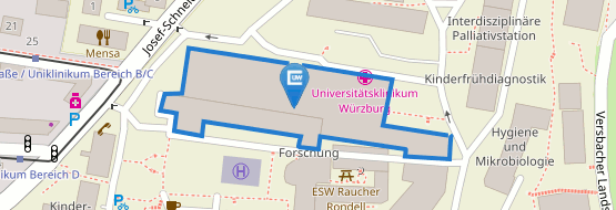
Structural Biology
Structural Biology
The following techniques are available to all research groups:
X-Ray Crystallography
Contact: Group Kisker, Group Schindelin
X-ray crystallography is the major technique for determining the three-dimensional structure of biological macromolecules. The crystals are usually grown by vapor diffusion, typically by the hanging drop method where a drop of protein solution is suspended over a reservoir containing buffer and precipitant. Water diffuses from the drop to the solution leaving the drop with optimal crystal growth conditions. The crystals should typically have minimum dimensions of 0.05 x 0.05 x 0.05 mm3. The crystals can comprise protein only, or a protein in a complex with nucleic acids (DNA or RNA) or another protein, or bound to a small molecule. The diffraction of X-rays through the evenly spaced crystal lattice produces the diffraction pattern. A series of diffraction patterns are taken while the crystal is rotated. By analyzing the observed X-ray diffraction patterns originating from the crystals and using sophisticated computational techniques, the three-dimensional structure of the macromolecule(s) in question is calculated.
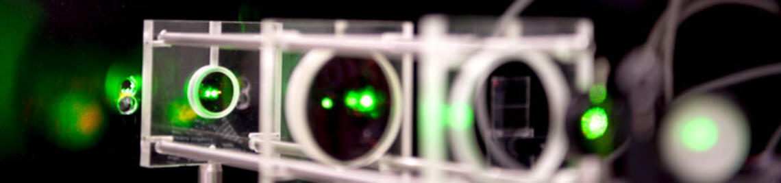
Cryo-Electron Microscopy:
Contact:
Prof. Böttcher Bettina (Professor for Biochemistry with the focus on Cryo-electron microscopy at the Department of Biochemistry of the University of Würzburg)
Tel.: +49 931 31-84193
E-Mail: bettina.boettcher@uni-wuerzburg.de
Biological objects such as complexes and viruses, are rapidly frozen in a thin film of vitrified buffer. These vitrified samples can be imaged directly in the electron microscope at temperatures between -170 and -180 °C. The images are phase contrast images and represent projections of the density of the object. Computer aided image processing enables combining many different projections of an object to a three-dimensional map. These maps can have resolutions in the order of 2-4 Å provided that thousands of projections of identical copies of the object can be coherently averaged and that the micrographs contain high-resolution information.
Typical objects for structure determination by electron cryo microscopy are complexes with more than 150 kDa and a thickness of less than 100 nm.