University of Würzburg strengthens biomedical microscopy
03/26/2020On 23th March 2020, Professor Katrin Heinze officially started her new “Chair of Molecular Microscopy" at the Medical Faculty of the Julius Maximilians University (JMU) Würzburg, Germany. The physicist will boost the development of precise microscopy methods for biomedical imaging and spectroscopy.
The visualization of spatial and temporal processes in the living organism expands our understanding of physiological and pathophysiological processes in the body. Modern imaging makes it possible to visualize individual molecules and their interactions in a living organism, as well as to investigate specifics of individual cells or the structure of entire organs or tumors.
Expansion of biomedical microscopy
Prof Katrin Heinze uses a variety of physical tricks and ideas from materials science. Her repertoire extends to the construction of microscope prototypes, special sample preparations and nano-coatings to quantitative 3D image analysis with customized evaluation pipelines. The establishment of the new Chair of Molecular Microscopy will strengthen and expand biomedical microscopy at the Rudolf Virchow Center for Experimental Biomedicine (RVZ) at the University of Würzburg, especially with extended premises and additional investment funds.
Numerous collaborations with both clinical and basic research colleagues, as well as the involvement in three ongoing DFG Transregio Collaborative Research Centers reflect the accuracy of her methods. “It is important to me to allow a wide range of questions benefit from our fluorescence techniques. We therefore plan to further develop our current methods (from the single molecule to the whole organ), with particular attention to fast, high-resolution procedures that can be combined with other medical imaging techniques or can be used to complement each other,” emphasizes Heinze.
Investigating molecular mechanisms in living cells and organs
Fluorescence-based imaging is a fascinating and indispensable tool in scientific research. "In my group, microscopy and image analysis methods are being developed for biomedical research with the aim of being able to investigate molecular complexes in living cells or whole organs. For example, we combine concepts of two-photon and light sheet microscopy with computer simulations, or high-resolution fluorescence microscopy with materials research techniques. Our latest development involves developing and manufacturing nano-coated cover glasses for super high resolution. Our biocompatible metal-dielectric coatings act as an 'amplifier' for various fluorescence applications and enable faster and more precise imaging," explains Heinze.
As this minimally invasive method is suitable for the analysis of living cells, the scientist expects, for example, fascinating and detailed insights into spatial and temporal distribution patterns of membrane receptors that would normally be hardly accessible using fluorescence methods.
Person
Katrin Heinze holds a doctorate in physics and has been enthusiastic about fluorescence techniques since her studies. Since then, her research has focused on the development of such processes with the highest temporal and spatial resolution. After completing her doctoral thesis on correlation methods in living cells in Petra Schwille's laboratory, which was awarded with the Otto Hahn Medal by the Max Planck Society, Katrin Heinze spent her postdoc time in Dresden (Germany) and then at McGill University in Montreal (Canada) in Paul Wiseman's laboratory, where she successfully devoted herself to novel lithographic and imaging techniques. Back in Europe end of 2006, she headed the “Optical Engineering” department at the Institute of Molecular Pathology in Vienna, before starting her own research group at RVZ in Würzburg (Germany) in 2011, with the aim of visualizing and analyzing disease-related processes as well as the architecture of tissues and biomolecular complexes.
Contact
Prof. Dr. Katrin Heinze (Rudolf Virchow Center, University of Würzburg)
Tel. +49 (0)931 31 84214, katrin.heinze@virchow.uni-wuerzburg.de
Dr. Daniela Diefenbacher (Press Office, Rudolf Virchow Center)
Tel. +49 (0)931 3188631, daniela.diefenbacher@uni-wuerzburg.de
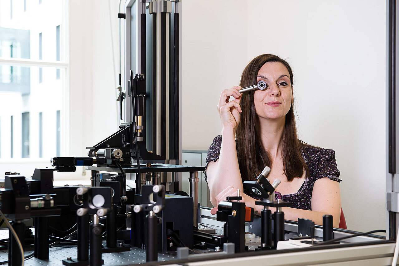
Photo
Prof Katrin Heinze, Rudolf Virchow Center, University of Würzburg (Foto: bettinaflitner.de)