Cutting-edge microscope for North Bavaria
01/17/2018This year, one of the world's most powerful electron microscopes will start operation at the University of Würzburg, providing images of biological molecules of unparalleled quality.
According to the manufacturer FEI, the Titan Krios is the most powerful and most flexible electron microscope in the world to provide high-resolution images of biological samples - both in 2D and 3D mode.
The Titan is presently being installed at the University of Würzburg. Last year, the German Research Foundation approved the corresponding application and released some EUR 3.8 million for the purchase. In addition to researchers from Würzburg, the microscope will also allow scientists from the universities in Bayreuth, Erlangen and Regensburg to take structural images of biological samples smaller than one millionths of a millimetre.
Recording images at minus 180 degrees Celsius
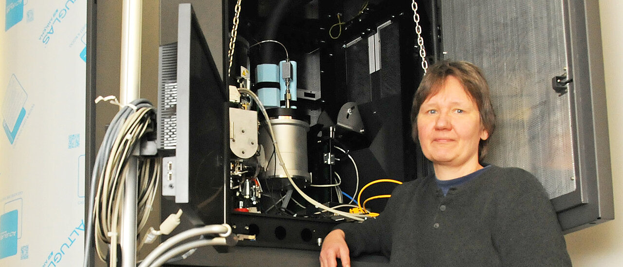
Bettina Böttcher is responsible for the microscope. The scientist has been a professor at the Department of Biochemistry of the University of Würzburg since August 2016. Her focus is the so-called "cryo-electron microscopy". This technology is characterized by extremely low temperatures of down to minus 180 degrees Celsius and a resolution at the atomic scale. It enables studying rapid-frozen biological molecules and complexes in solution and reconstructing their three-dimensional structure.
The microscope accelerates the electrons it uses to "scan" the test specimens at a voltage of 300,000 V. Once in operation, it will produce up to two terabytes of image data each day. But since it may take several thousand images to provide a 3D model of a molecule, a single analysis can last between two and four days.
Resolution at the atomic scale
"The device allows us to automatically record high-resolution data during several days of non-stop operation. Depending on the specimen, the microscope yields structural data of two to four ångström," Professor Bettina Böttcher explains. An ångström is equal to 0.1 nanometre. It is the unit of length for distances of atoms in crystal structures and the lengths of bonds in molecules.
Moreover, the electron microscope is capable of automatically processing up to 12 samples in parallel while maintaining the vacuum. But the scientists also appreciate another property of the electron microscope: "We can recover the samples free from contamination to study them further subsequently," Bettina Böttcher explains.
Specific requirements for the installation environment
The people in charge at the Rudolf Virchow Center found a suitable room to install the machine. This room has to meet a number of special requirements: For the microscope to deliver high-quality images, it must be protected against vibration, sound and moisture and screened against electromagnetic radiation.
Having arrived in Würzburg in late 2017, the microscope is currently being set up. If everything proceeds as scheduled, Bettina Böttcher plans to start routine operation of the microscope in the next months.
Contact
Prof. Dr. Bettina Böttcher, Professor of Biochemistry with a focus on cryo-electron microscopy,
T: +49 931 31-84193, bettina.boettcher@uni-wuerzburg.de
Additional images
.")