Correspondence between cells is strictly regulated
02/02/2018In order to communicate, animal cells send each other messages in a bottle. Researchers from Würzburg have now been able to demonstrate how this process is regulated.

Animal cells are avid pen pals: they send each other membrane vesicles filled with signaling molecules in order to communicate. Scientists from the Rudolf Virchow Center for Experimental Biomedicine at the University of Würzburg have determined how this process is regulated. Their results were recently published in the scientific journal, the Proceedings of the National Academy of Sciences (PNAS). In the long run, medicine could also benefit from their findings.
Interaction between cells
A thin barrier called a membrane surrounds every animal cell and separates it from its environment. The membrane consists of a bilayer of fat-like molecules called lipids. Cells can use this lipid membrane to wrap information and send it to other cells - much like a letter in an envelope. Cells bud out a small membrane bubble called a vesicle from their surface and these vesicles are filled with or covered in signaling molecules. This form of communication enables cells in the same neighborhood to coordinate their behavior.
Researchers have long known about the existence of these intercellular messages in a bottle. How exactly their release is regulated was largely unknown. Scientists led by Ann Wehman from the Rudolf Virchow Center of the University of Würzburg have made a significant advance in clarifying this question. "We were able to identify a particular enzyme that plays a key role in the formation of the vesicles, the TAT-5 flippase," explains Katharina Beer, who is a PhD student in the Wehman lab.
TAT-5 prevents lipids from flipping out
The membrane bilayer consists of an inner and an outer lipid layer. Many different molecules are embedded in both layers, some of which are specific to a particular side. An example of this asymmetry is the distribution of the so-called PE lipids. They usually stay in the inner membrane. The TAT-5 flippase ensures that this distribution does not change: as soon as PE molecules move into the outer membrane, the enzyme "flips" the misrouted molecules back to the inner layer.
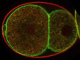
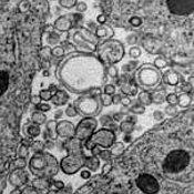
But what happens when the TAT-5 flippase fails? In this case, PE lipids accumulate in the outer membrane. At the same time, the cell begins to release numerous vesicles. "Vesicle release is apparently initiated by the redistribution of the PE molecules," explains Katharina Beer.
The scientists also analyzed various genes involved in the formation of the vesicles in nematode worms. They identified eight genes that ensure the correct functioning of the TAT-5 flippase. One of the eight seems to regulate the activity of the "asymmetry enzyme" directly.
The others ensure that the flippase reaches the cell membrane in order to perform its task. "We inhibited these genes in worms - either one or two at a time," says Beer. "As a result, we observed a massive production of vesicles."
Too much of a good thing
Due to the large number of membrane vesicles released, the embryonic development of the worms was severely disrupted. "It's as if the cells were walking on marbles," says Wehman, " and were not able to make any progress." For example, the intestine develops on the surface in some embryos instead of inside the body.
The research group will now explore why the redistribution of PE lipids has such a serious impact on vesicle production. Wehman recently raised half a million Euros in research funding from the German Research Foundation (DFG) for this project.
To explore these processes in more detail is also relevant from a medical point of view. It is known that cancer cells release membrane vesicles in large quantities and that these messengers promote tumor growth. Immune cells use the intercellular mail service to inform each other about an infection. Communication via extracellular vesicles also plays a role in the formation of blood clots, which can impact heart attacks or stroke.
The findings of the Würzburg researchers provide a foundation to gain a better understanding of these medically relevant cellular processes. In the long run, they could help to improve the treatment of certain diseases.
Publication
Katharina B. Beer, Jennifer Rivas-Castillo, Kenneth Kuhn, Gholamreza Fazeli, Birgit Karmann, Jeremy F. Nance, Christian Stigloher und Ann M. Wehman: Extracellular vesicle budding is inhibited by redundant regulators of TAT-5 flippase localization and phospholipid asymmetry; PNAS, Januar 2018; DOI: 10.1073/pnas.1714085115
People
Dr. Ann Wehman moved to Germany in 2013 to lead a research group in the Rudolf Virchow Center for Experimental Biomedicine at the University of Würzburg.
Katharina Beer is a PhD student in the research group of Dr. Ann Wehman.
Webpage AG Wehman
Kontakt
Katharina Beer (Wehman Group, Rudolf Virchow Center)
Tel. +49 931 3188955 , katharina.beer1@uni-wuerzburg.de
Dr. Daniela Diefenbacher (Public Science Center, Rudolf Virchow Center)
Tel. +49 931 3188631, daniela.diefenbacher@uni-wuerzburg.de


