SEM (JEOL JSM-7100F) and XRM (XRM II)
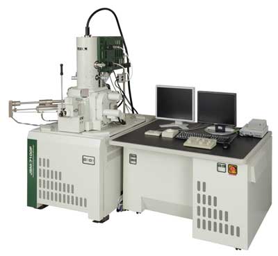
Scanning Electron Microscope (JEOL JSM-7100F) equipped with a X-ray microscope (XRM II)

Specifications
Scanning Electron Microscop (JEOL JSM-7100):
- SEI Resolution: 1.2 nm (30 kV)
- Magnification: 10 - 1 000 000
- Accelerating Voltage: 0.2 - 30 kV
- Probe Current: 1 pA - 400 nA
- Electron Gun: In-lens Schottky field emission gun
- A state of the art scanning electron microscope
X-ray microscope (XRM II):
- Source: Customized JEOL JSM-7100F (Vacc 30 kV, Imax 400 nA)
- Reflection Target: Nanostructured molybdenum and tungsten
- Detector: Photon-counting direct-converting X-ray detector with 768x512 pixels
- Magnification: up to 1000
- Resolution: down to 50 nm
- Fully 3D imaging possible through piezo-powered rotational axis
Contact person | Location |
|---|---|
|
Examples
Setup of an electron probe micro analyzer for highest resolution radioscopy
R. Hanke, F. Nachtrab, S. Burtzlaff, V. Voland, N. Uhlmann, F. Porsch and W. Johansson
Nuclear Instruments and Methods in Physics Research 1, 173 (2009)
Laboratory X-ray microscopy with a nano-focus X-ray source
F. Nachtrab, T. Ebensperger, B. Schummer, F. Sukowski and R. Hanke
Journal of Instrumentation 6, C11017 (2011)
A laboratory X-ray microscopy setup using a field emission electron source and micro-structured reflection targets
P. Stahlhut, T. Ebensperger, S. Zabler, R. Hanke
Nuclear Instruments and Methods in Physics Research Section B 324, 4–10 (2014)


