High-content 3D-Imaging and image-based modelling
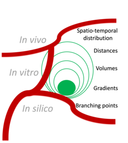
In biomedical and translational imaging, we have several challanges to overcome. Besides resolution and speed, the „Field of view“ that we can cover is of crucial importance. Some findings are only meaningful if put into the tissue context, ideally covering the whole organ. Thus, fluorescence imaging can uniquely provide insights into the intricate mechanisms of organ function and disease progression. By developing imaging pipelines and tools that allow for high-resolution imaging of these interactions, we contribute to a better understanding of the underlying biology. We work on our concepts and respective imaging pipelines with focus in cell-vessel interactions in murine organs. Our comprehensive approach starts from the hardware aspects of microscopes and extending to scripting for quantitative data analysis to create a complete imaging solution from data acquisition to interpretation. In the past years we have contributed to a large number of biomedical projects and tailored and built the imaging framework starting with the microscope „hardware“ and ending with scripts for quantitative data analysis.
Selected topics are
- Ischemic stroke (in collaboration with Prof. David Stegner)
- Megakaryopoiesis and Thrombocytopenia (in collaboration with Prof. David Stegner)
- Thromboinflammation (in collaboration with Profs David Stegner and Bernhard Nieswandt)
- Fungal infections in the lung (in collaboration with Prof. Andreas Beilhack)
- Tumor therapy (in collaboration with Dr. Erik Henke)
Given the rapid advancements in imaging technologies, we believe our work has not only implications for basic research but also for potential clinical applications in future. Imaging techniques and tools developed in preclinical studies involving murine models can often pave the way for improved diagnostic and therapeutic strategies in human medicine.


