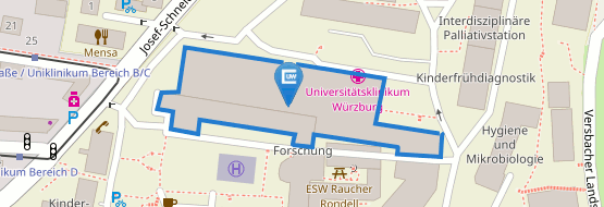
OUR RESEARCH
WELCOME TO THE HEINZE LAB

Fluorescence approaches have become an integral part of life sciences by providing researchers with a powerful tool to visualize cellular, subcellular and even molecular structures and conformations. High-resolution translational imaging with a focus on tissue context and dynamics provides us with a comprehensive understanding of biological systems, ultimately leading to advances in our knowledge of health and disease.
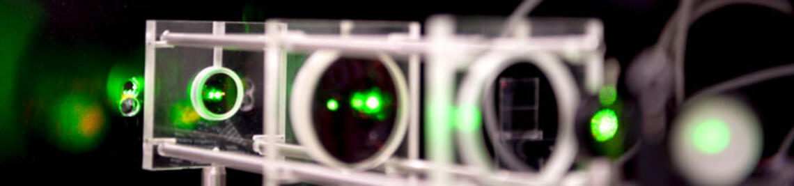
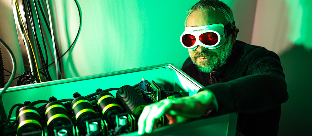
Fluorescence approaches in science resemble captivating black light theater on stage. By harnessing the power of fluorescence, we can see what lights up in biological systems. The remarkable selectivity, subcellular resolution, and speed offered by fluorescence microscopy allow us to unravel the intricate architecture of organs and cells, as well as the underlying dynamics of molecules. Together with our partners from universities and clinics, we address complex scientific inquiries that can only be tackled through multi-faceted approaches. This is why we embark on a journey that spans from designing customized microscopes to developing specialized imaging pipelines and crafting individual analysis scripts.
Imaging across scales is our mission.
Fluorescence microscopy ranging from whole organs to individual molecules is a challenging task. While resolution plays a crucial role in translational imaging, it is equally important to consider the tissue context surrounding the subcellular architecture being investigated, as well as the dynamics of the living system under study. By understanding both the spatial organization and the functional aspects of biological structures, we gain a more comprehensive understanding of processes underlying health and disease.
Taking a journey through any cell, organ and organism is our vision.
The vision of our imaging approach is to take a journey through an organ or cell to make the connection between architecture, function and dysfunction. We want to develop tools that allow seamless zooming in and out, enabling visualization from the level of single molecules to that of entire organs. By segmenting three-dimensional images, we can create biological templates or "digital playgrounds" to test complex hypotheses in silico to better guide our in vivo experiments. Together with augmented reality tools of our partners from informatics, such virtual experiments and simulations may provide a platform in itself like a “holodeck” to test hypotheses in future and gain insights into the biological players involved.
Methods, methods, methods
- Light-sheet fluorescence microscopy
- Video microscopy
- Confocal Microscopy
- Expansion Microscopy
- Single molecule localization microscopy
- Time-resolved fluorescence (FLIM, FCS, Anisotropy)
- Metal-enhanced super-resolution
- Image reconstruction, segmentation and quantitative feature extraction